Authors of the top accepted contributed papers that describe medical devices with commercial potential will be giving five minute pitches to a panel of leading medical technology innovators. The top three presentations will be chosen to win one of three cash prizes.
The Judge's Panel will base their decision upon four factors:
1) Quality of clinical need statement (problem)
2) Technical soundness of research (solution)
3) Presentation Quality
4) Fundability
Winners will be announced during lunch following the competition on Wednesday.
Judges for the 2024 Competition Include:
Paul Rothweiler, Bakken Medical Devices Center
Bill Betten, S3 Connected Health
Dawn Bardot, Abiomed
Aghogho Ekpruke, Medtronic
Del Lawson, Solventum
Kate Taylor, Boston Scientific
Tim Tripp, ATP-Bio
Mark Wehde, Mayo
Design of a New Pleural Biopsy Device for Improved Procedural Efficacy (Grand Prize Winner)
Pleural biopsies are challenging procedures that rely heavily on the skill of the performing clinician as there is little to guide them other than the feel of the needle within the patient. This is especially true in resource-poor settings as the cost and availability of external imaging equipment (such as computed tomography, ultrasound scanners, or thoracoscopes), and the increased skill needed to use the equipment prohibits their regular usage during these procedures. As a result, basic cutting needle biopsies have high risks of causing damage to the lung when the needle is inserted past the pleural space, or low diagnostic rates as pleural tissue is often not included in the biopsies. However, despite these drawbacks, pleural biopsies are performed regularly, most notably for diagnosing tuberculous pleurisy in low- or middle-income countries. This paper proposes a new cutting needle design to firstly improve the yield of pleural tissue in the biopsies, and secondly, allow for multiple biopsies to be collected with a single insertion, minimising the risks usually associated with each needle insertion. Preliminary verification of the biopsy mechanism shows that multiple, separate, adequately sized biopsies are possible with this design. Watch the pitch.
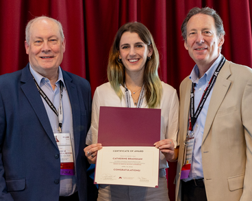
Catherine Bradshaw, PhD
Biomedical Engineering Candidate, University of Cape Town
Catherine is a final-year PhD candidate in biomedical engineering at the University of Cape Town, under the supervision of Profs Sudesh Sivarasu and Keertan Dheda. She is a member of UCT Medtech and is committed to bringing low-cost medical interventions made in South Africa to South Africa and the rest of the African continent. Her research embodies the intersection of innovation, patient care, and advanced medical technologies and focuses on improving diagnostic methods through smart technology. Before her PhD in Biomedical Engineering, Catherine completed an undergraduate mechatronic engineering degree at the University of Cape Town which built on her love of all things electronic.
A Flexible Patella Fracture Device for Increased Anterior Cortical Compression (2nd Place Winner)
Fractures of the patella can be challenging to treat surgically. The current standard of tension band wiring is susceptible to wire loosening and fracture displacement. In this work we introduce a novel concept involving a flexible patella fixation device which leverages pre-strain to deliver interfragmentary compression concentrated anteriorly. Finite element analysis provides a preliminary exploration of the concept, demonstrating that two different flexible patella plates can provide increased anterior compression both alone and when paired with lag screws. Watch the pitch.
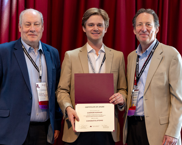
Connor Huxman
PhD Candidate, Mechanical Engineering, Penn State University
Connor has been designing orthopaedic devices in industry and academia for the last five years. This has included working as an R&D engineer at a start-up on the design and regulatory approval of spinal fusion implants, as well as academic research on the design of new devices for fracture fixation. He received his B.Sc. in Biomedical Engineering in 2020 from the University of Waterloo (Ontario, Canada), his M.S. in Engineering Design in 2022 from Penn State University, and is currently working towards his PhD in Mechanical Engineering at Penn State. Connor is passionate about the intersection of orthopaedic implant design, regulatory strategy, and surgeon adoption. He is currently developing a new technology for delivering controlled micromotion to long bone fractures with the goal of facilitating faster and stronger bone healing.
Scan, Aim, Go: Compact Brain Drill Guide for Accelerating Minimally-Invasive Neurosurgeries (3rd Place Winner)
Workflows for MRI-guided minimally-invasive neurosurgeries are often time-consuming and complex. Many minimally-invasive interventions utilize magnetic resonance imaging (MRI) to noninvasively visualize internal anatomy and pathologies. This, in conjunction with external trajectory guides, can be used to internally position devices to perform complex surgeries with minimal disruption to the patients’ healthy anatomy.
These trajectory guides are typically rigidly attached to the skull and feature adjustable channels through which drills, needles, and /or catheters may be introduced. Trajectories are oriented by iterating between imaging and manipulation of device settings. MRI scans, while offering a great deal of valuable anatomic information, are slow to acquire. Scans of a sufficient resolution for neurosurgery can take on the order of 10 minutes to acquire, depending on field of view. Noniterative approaches could reduce complexity and anesthesia time.
This work describes efforts to create and validate a new trajectory guide that enables faster, accurate trajectory guidance in minimally-invasive neurosurgeries. Using new hardware and software, a single scan approach was used to perform drill guidance and device insertion on phantoms and cadaver heads.
The proposed methodology accurately guided needles to targets within phantoms and human cadaver brains using a single targeting scan. The initial design produced a radial error of 1.4±0.8mm in phantoms and 1.5±0.8mm in cadaver brains.
The proposed device and software accelerate trajectory guidance in minimally-invasive neurosurgeries by reducing the number of acquired scans and procedural steps. This in turn minimizes time under anesthesia. Watch the pitch.
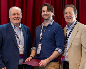
Thomas Lilieholm
University of Wisconsin at Madison, Medical Physics
Tom Lilieholm, MS, is in the fifth and final year of his PhD studies in Medical Physics at the University of Wisconsin-Madison under the supervision of Dr. Walter F. Block. Here, he works with the university’s Interventional MRI team developing workflows and tools for MR-guided minimally invasive interventions. As a member of the UW Biotechnology Training Program grant, Tom also meets with local research commercialization resources to evaluate innovations developed within the university. His research interests include machine learning, image analysis, and quantitative MRI, with a focus on procedures like prostate cryoablation, intracerebral hemorrhage evacuation, convection enhanced delivery, and the placement of deep brain stimulators.
Prior to UW, Tom received his Bachelor’s in physics from the University of Texas at Austin, where he also earned a minor in business through the Red McCombs Summer Institute program and researched the physics of bacterial biofilm adhesion within UT’s Center for Nonlinear Dynamics.
A Novel Tool for Application of Bone Wax in Neurotologic and Skull Base Surgery
Surgeries of the ear and lateral skull base frequently require the use of bone wax to seal off small vessels and to repair or prevent leakage of cerebrospinal fluid. Application of bone wax to the hard, wet, and irregular surface of the bone is challenging with conventional instruments due to the tendency of the wax to slip off the bone and the narrow surgical corridors of the lateral skull base. Here we present a novel surgical tool for bone wax application, comprised of a rigid core with a coating of softer material over the ends. We tested several combinations of 3D printed materials in cadaveric temporal bones simulating surgical conditions. The tool was superior to conventional methods of bone wax application for all material combinations tested. Further studies into optimal materials and additional applications are planned. Watch the pitch.
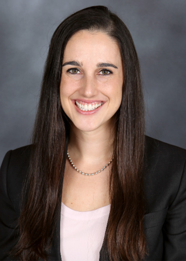
Miriam Smetak
Fellow, Otology & Neurotology, Washington University in St. Louis
Miriam Smetak is a Fellow in Otology, Neurotology and Skull Base Surgery at Washington University in St. Louis. She completed her M.D. and a concurrent M.S. in Biomedical Engineering at the University of Minnesota, followed by residency in Otolaryngology-Head & Neck Surgery at Vanderbilt University. Miriam leverages her engineering background to solve challenges in the operating room, with a particular focus on human factors and latent sources of error.
Development of a novel intra-abdominal catheter placement device for use in telesurgery
Telesurgery applications in laparoscopic surgeries are limited by the need for a surgeon to manually insert the trocar for access to the abdominal cavity. This feasibility study aims to solve this by capitalizing on the biomechanical properties of the abdominal wall to introduce the Robotic Automated Peritoneal Insertion Device (RAPID), a novel mechanism employing a purely mechanical indication system for accessing the abdominal cavity. The RAPID leverages pressure differences between layers in the abdominal wall and the abdominal cavity to indicate entry.
In a previous study, optimal pressure parameters for the RAPID were determined through experiments conducted on cadaver and porcine models. In the present work, the RAPID utilizes this optimal pressure in an entry indication system that is validated in live porcine models. The system operated correctly in 19 out of 21 placements, affirming the feasibility of the entry indication system. This shows that the RAPID can accurately indicate entry of the trocar into the abdominal cavity. This groundbreaking advancement has the potential to automate initial and subsequent trocar insertions, thus removing one more constraint on laparoscopic telesurgery. Watch the pitch.
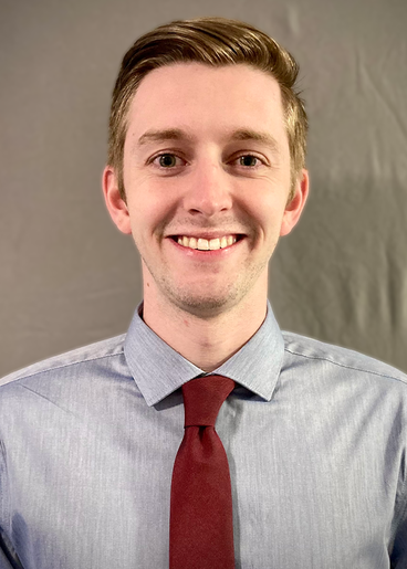
Gregory Hirst
Undergraduate, Mechanical Engineering, Brigham Young University
Greg Hirst is a senior at Brigham Young University, diving into research at the Terry Research Lab where he explores tissue characterization and helps develop innovative medical devices. He's excited to begin his master’s studies at BYU in fall 2024, eager to explore mechanobiology and continue his journey in cutting-edge biomedical research.
Flexible Precision: Design and Testing of a Snake-Inspired Robotic Arm for Large Organ and Tissue Retraction during Robot-Assisted Surgery
In the realm of robot-assisted surgery, maneuverability in confined spaces remains a critical challenge. This paper presents the development of a snake-like robotic arm designed specifically for large organ and tissue retraction during minimally invasive surgeries. Emulating the flexible movements of a snake, the robotic arm aims to push the boundaries of current surgical robots. Key objectives include compatibility with existing minimally invasive ports, capacity to hold up to 2 pounds of tissue without deformation, axial stability under 10N force at full extension, compatibility with articulate-by-wire robotic systems, and a minimum of 4 degrees of freedom.
The design and construction involve three segments: the base attachment, middle links, and the end manipulator. Finite Element Analysis (FEA) validates the design and demonstrates the device’s ability to handle up to 10N before the onset of deformation. The arm, manufactured using stereolithography printing, handled 2 lbs (~10N) of distal-segment load thus providing real-world evidence to reinforce the FEA data. The actuation base, powered by high-torque servo motors controlled by a separate control module, provides precise and responsive movement. Extensive testing, both in simulated computational environments and physical stress tests, demonstrates the arm's proficiency in handling surgical tasks within minimally invasive procedures. Watch the pitch.

Basil Alias
Texas A&M School of Engineering Medicine
Basil Alias is an M.D. and M.Eng candidate at Texas A&M University, School of Engineering Medicine (ENMED). With a background in biomedical engineering and experience in spinal instrument and implant development, has led him to learn about and innovate in medicine. In this endeavor, in partnership with the Houston Methodist Research Institute and with support from the Brown, Smith, and Raymond ENMED Capstone Program, he hopes to make a lasting impact on healthcare through innovation in surgery and medical visualization.
In the future, Basil plans to use his experience in engineering to bolster his medical expertise. He hopes to specialize in otolaryngology, to make an impact on those suffering from head and neck diseases and disorders. In his commitment to medicine, he has been able to develop, implement, and generate innovative devices and technologies, such as software for facial paralysis, instrumentation for facial reconstructive procedures, and more, that are used clinically, and he hopes to continue this trend throughout residency, and for the rest of his career in medicine.

Raaghav Bageshwar
Texas A&M School of Engineering Medicine
Raaghav Bageshwar stands at the confluence of engineering and medicine as an M.D. and M.Eng. candidate at Texas A&M School of Engineering Medicine (EnMed). With a robust foundation in Biomedical Engineering, Raaghav's drive for innovation propels him to collaborate with a classmate and friend on a groundbreaking project: a snake-like robotic arm intended for minimally invasive surgeries. This endeavor, in partnership with the Houston Methodist Research Institute and supported by the Brown, Smith, and Raymond EnMed Capstone Program, exemplifies his goal to enhance medical procedures through the application of engineering innovation and medical knowledge.
Looking toward the future, Raaghav plans to merge his biomedical engineering background with his growing medical expertise. He aspires to specialize in internal medicine, followed by a cardiology fellowship, aiming to make significant strides in the cardiology space. This path reflects Raaghav’s dedication to using his unique skill set for cardiac care advancements. Through his commitment to healthcare innovation, Raaghav aims to significantly impact patient care, paving the way for new paradigms in the integration of engineering advancements within the healthcare industry.
Development of a Self-Powered Sensory Neuroprosthesis with Pneumatic Actuators
Peripheral neuropathy (PN) results from damage to the nerves in the peripheral nervous system [1] and can cause loss of feeling or numbness in the hands and feet [2]. This leads to several challenges, including difficulty walking. Our goal was to create a device that gives stimulus during gait to a portion of the limb that still has sensation and ultimately reduces the chance of trips and falls. Additional constraints were placed on the system being cost-effective and easy to use. To do this, we have composed a device using a closed, human-actuated air bladder system. An input air bladder underfoot compresses during walking, which in turn, inflates an air bladder on the leg that stimulates sensory nerves. To test the device, an Instron was used to apply a load to the input bladder, and the force generated by the output bladder was measured on a mannequin leg. Four devices were evaluated in total, with two different output bladder designs and two different fluid connection systems between the bladders. It was found that both output bladder designs were effective and the connection system consisting of polyurethane tubing was superior in translating the input force onto the leg. This design also had the fastest force release, which could prove beneficial during the naturally cyclical process of walking. Watch the pitch.
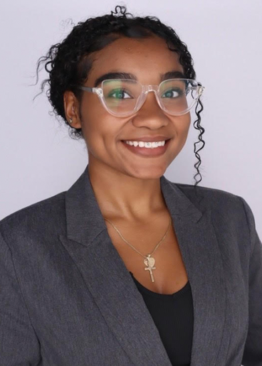
Bianca Campos
Student, Biomedical Engineering, Virginia Tech
I'm Bianca Campos, a dedicated undergraduate student studying Biomedical Engineering at Virginia Tech. With a passion for healthcare innovation, I've gained hands-on research experience analyzing data and developing medical devices. I'm committed to making a positive impact in the field and beyond.
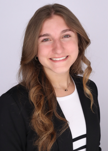
Mollie Schoeppner
Undergraduate Student, Biomedical Engineering, Virginia Tech
I'm Mollie Schoeppner, an undergraduate student studying Biomedical Engineering at Virginia Tech. I'm committed to merging engineering with the healthcare and medical device field. I have been gaining research experience designing medical devices and evaluating data. I hope to continue research and have a valuable contribution to the field.
Non-Invasive, Portable Optical Imaging Device for Early Detection of Melanoma in Aging Populations using Diffuse Reflectance Spectroscopy (DMD2024-1001)

Jeff J.H. Kim
MD/PhD Student, Biomedical Engineering, University of Illinois College of Medicine
Skin cancer is the most prevalent form of cancer, with melanoma being the most deadly form. Early detection is crucial for effective treatment, yet it remains challenging, as accurate diagnosis often requires expert medical knowledge. This study presents a novel, portable, and non-invasive optical imaging device designed for early detection of melanoma in clinical and non-clinical settings. By leveraging diffuse reflectance spectroscopy and incorporating a new 'Z criterion' into the established ABCD rule (Asymmetry, Border, Color, Diameter), the device offers an enhanced assessment. The preliminary study on a cohort of elderly patients demonstrated the device's feasibility in detecting melanoma, showing significant promise for application in portable diagnostics and at-home care. Jeff was unable to compete in person at the last minute.